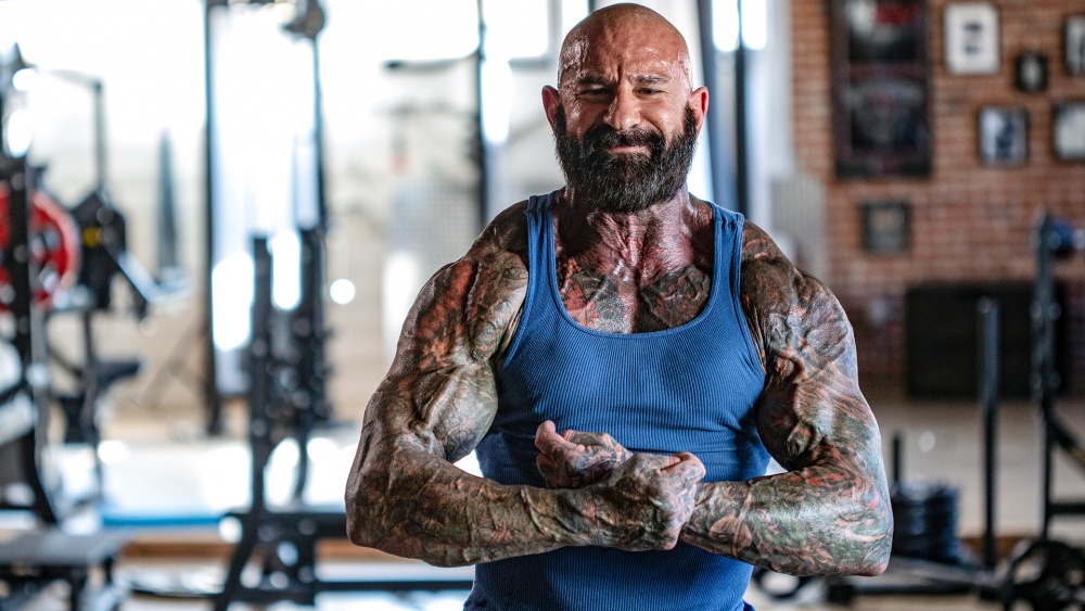
Beginner's Guide to Musculature
Learn the how and why of resistance training with this walkthrough of all the major muscle groups involved in training, how they work, and which exercises can be used to train them.
Learn the how and why of resistance training with this walkthrough of all the major muscle groups involved in training, how they work, and which exercises can be used to train them.

Obviously, we use our muscles on a daily basis. But when it comes to strengthening them, increasing their size, and conditioning them to endure more exertion for longer, exercise and weight training in particular are the path to improved musculature. In order to know how your muscles function during exercise, it’s important to understand their basic structure and function.
On the largest scale, skeletal muscle tissue is divided up into muscle groups with which most people are familiar. The pectoral muscles, for example, are the muscles of the chest, responsible for pressing movements like the bench press and push-ups. These large bundles of muscle tissue are connected to the bones of the body by tendons. Contraction of the muscles pulls on the connected bones, which facilitates movement of those bones.
Muscle tissue is composed of bundles of what are known as fascicles, which are themselves bundles of individual muscle fibers.
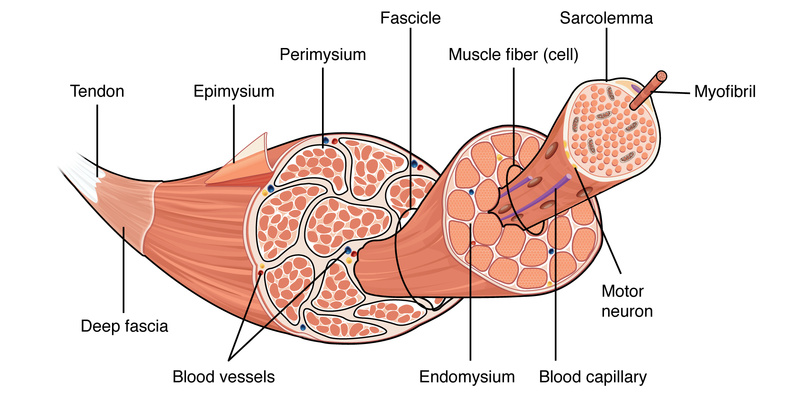
Muscle fibers, also called myocytes, are the unique cells of the body that make movement possible. Each muscle fiber contains multiple nuclei, the number of which is increased by muscle protein synthesis induced by exercise. This is one way in which muscles are thought to grow.
These muscle cells are filled with a fluid called sarcoplasm which, in addition to providing a medium through which the muscle cell’s exchange of nutrients and other bodily chemicals can travel, contains glycogen—stored glucose which the muscle tissue uses for the production of energy—as well as myoglobin, which stores oxygen also used in energy production inside the muscle.
Within these specialized cells are bundles of myofibrils, which are where muscle contraction actually takes place.
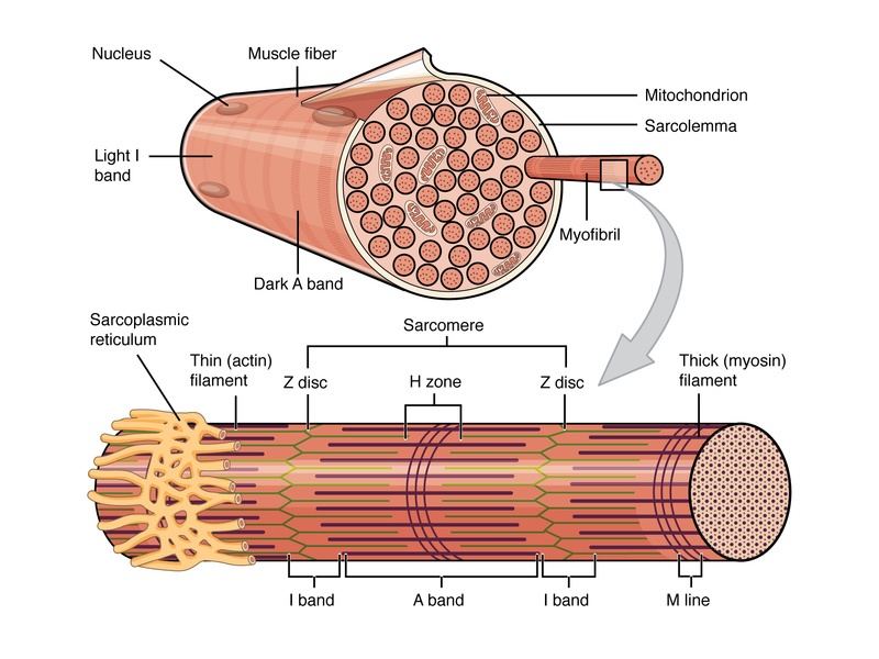
Myofibrils inside the muscle cells are composed of sarcomeres—long tubes of proteins that interact with each other to make muscle contraction possible. Many different protein strands make up each sarcomere, and each muscle cell may contain hundreds of thousands of sarcomeres. The most important protein strands or filaments are myosin and actin. During the concentric portion of a movement, myosin and actin attach to each other, shortening the sarcomere. This shortening, performed by thousands of myofibrils, contracts the overall muscle tissue of which they are a part.
During the eccentric or negative portion of a movement, the sarcomeres must lengthen again. The myosin filaments release the actin filaments to make this possible. When muscle is lengthened while still under stress, as with heavy weight training, the actin is torn from the myosin. This damage and its subsequent repair are part of the process through which muscle mass is developed.
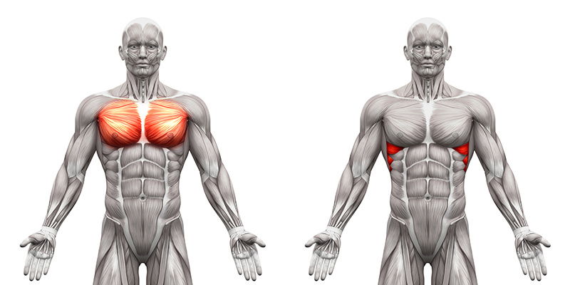
The chest refers primarily to the muscle group known as the pectoralis major. This consists of the upper pectoralis major and lower pectoralis major. The pectoralis muscles perform movements such as horizontal adduction of the upper arms, as in the dumbbell flye.
Basic, multi-joint exercises for the chest involve pressing movements, such as the bench press, incline dumbbell press, and push-up. Isolation exercises for the chest are flye-like exercises that involve movement of the arms without any change occurring at the elbow joint. Examples of isolation exercises for the chest include the dumbbell flye, cable crossover, and pec deck.
The upper and lower sections of the chest are hit differently by the various chest exercises. Therefore, the first order of importance when it comes to chest training is to ensure that you include exercises that target the upper, middle, and lower pectoralis.
Chest Muscle Structure
Chest Muscle Function
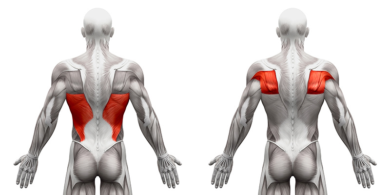
Back refers to the muscles that make up the backside of the torso. Although the term back refers mostly to the large latissimus dorsi muscles, or lats, that run from the upper arms all the way down to the buttocks, it can also include the teres major, the rhomboids, and even the middle and lower portions of the trapezius, because these muscles are often involved in performing exercises that are considered back exercises.
The two major types of lat exercises are the pulling exercises, which include pull-ups and pulldowns, and rowing exercises, which include bent-over barbell rows, T-bar rows, and seated cable rows.
Pull-up and pulldown exercises tend to concentrate more on the upper and outer lats as well as the teres major. Rowing exercises tend to concentrate more on the middle and lower lats as well as the rhomboids and middle trapezius muscles. Other types of lat exercises are the pullover and straight-arm pulldown.
The term back also refers to the musculature of the low back. The muscles in the lower back are those that support the spinal column and allow it to extend back, such as when you recline in a chair. These are deeper muscle fibers such as the spinal erectors, which include the longissimus thoracis, iliocostalis lumborum, and spinalis thoracis. Exercises that train the low back are back extension exercises and good mornings.
Back Muscle Structure
Back Muscle Function
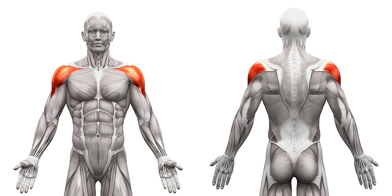
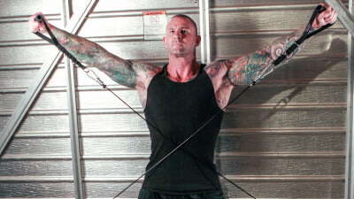
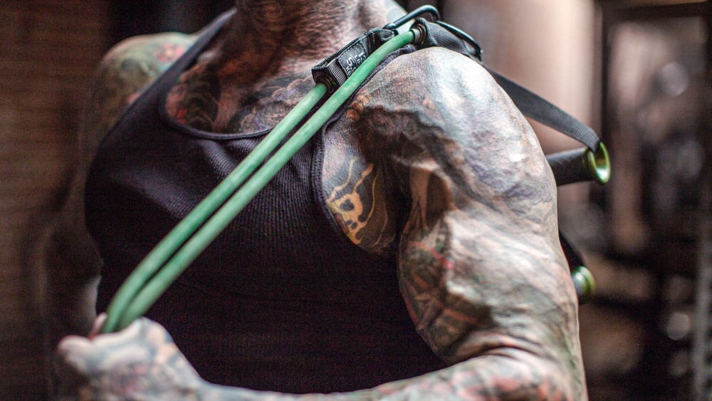
The shoulders refer to the deltoid muscles found on top of the upper arm. The deltoid is composed of three heads that originate on different points of the shoulder girdle but all converge on one common tendon that inserts on the humerus. The three heads are the anterior deltoid (front head), the middle deltoid, and the posterior deltoid (rear head).
Although these three heads work together to lift the upper arm at the shoulder joint, such as during the lateral raise, each head is stressed differently by different exercises. That is why it is important to structure shoulder workouts around basic multi-joint movements such as the shoulder press that hit all three heads, as well as isolation exercises such as front raise for the anterior head, lateral raise or upright row for the middle head, and rear deltoid raise for the posterior head.
Shoulder Muscle Structure
Shoulder Muscle Function
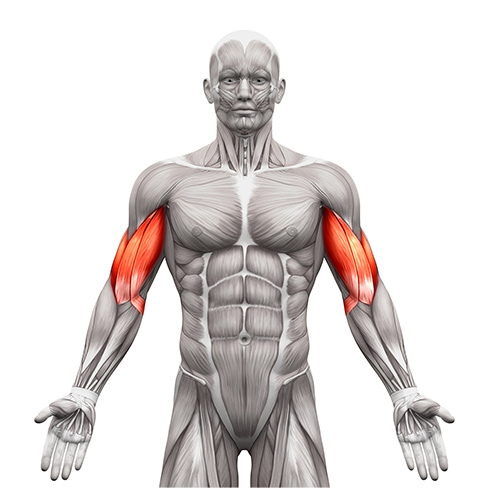
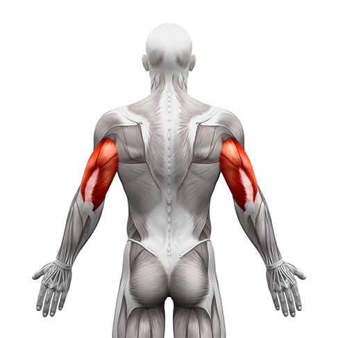
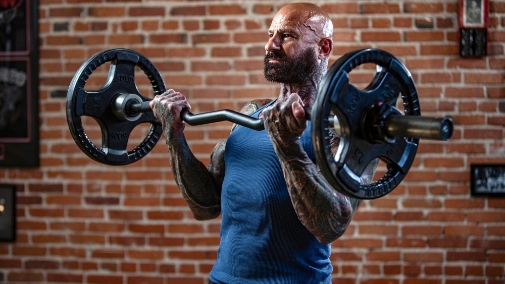
Biceps refers to two muscle heads that run down the front of the upper arm that are called the biceps brachii. The two heads are the long head (or outer head) and the short head (or inner head). The major difference between them is where each muscle attaches on the scapula. The tendon of the long head attaches farther back on the scapula than the short head. This is why they are referred to as long head and short head. Both biceps heads converge into one tendon near the elbow, and this attaches to the radius to cause flexion of the elbow when the muscles contract, such as when curling a dumbbell.
To flex the elbow, the biceps brachii receives help from the assistance muscle called the brachialis. This muscle lies underneath the biceps muscles and starts at the humerus and attaches to the ulna. The bulk of this muscle is lower than the bulk of the biceps muscle, which allows it to offer the most help during the first 30 degrees of elbow flexion. The brachialis is also strongly involved in elbow flexion when the hands maintain an overhand grip on the bar. The brachioradialis, although considered a forearm muscle, also helps at the initiation of elbow flexion when the hand is in a neutral position, such as during hammer curls.
Biceps Muscle Structure
Biceps Muscle Function
The triceps consist of three muscle heads that are on the back of the upper arm. The three heads of the triceps are the lateral head, long head, and medial head. Each head has a distinct attachment on the upper end, but they all meet at one common tendon that crosses the elbow and attaches on the ulna. Contracting the triceps results in extension at the elbow, such as the motion the arm makes when hammering.
The two types of triceps exercises are compound movements and isolation movements. Compound triceps exercises involve extension at the elbow and movement at the shoulder. These include close-grip bench presses and dips. Isolation triceps exercises involve just extension at the elbow with no other joint movement, such as dumbbell kickbacks.
Although every triceps exercise hits all three heads to some degree, certain ones are better than others at stressing the different heads because of the biomechanics involved. Because the long head of the triceps attaches to the scapula, it is more strongly contracted during exercises where the arms are brought overhead or in front of the body. This is because that action stretches the long head. Muscles contract the strongest when they are stretched to their longest length. Therefore, exercises that are done overhead such as overhead extensions best stress the long head of the triceps.
Exercises that place the arms in front of the body, such as lying triceps extensions also hit the long head to some degree. Extensions that are done with the arms at the sides of the torso while holding a neutral or overhand grip—such as triceps pressdowns and dumbbell kickbacks—best target the lateral triceps head. The same exercises done with an underhand grip seem to stress the medial head.
Triceps Muscle Structure
Triceps Muscle Function
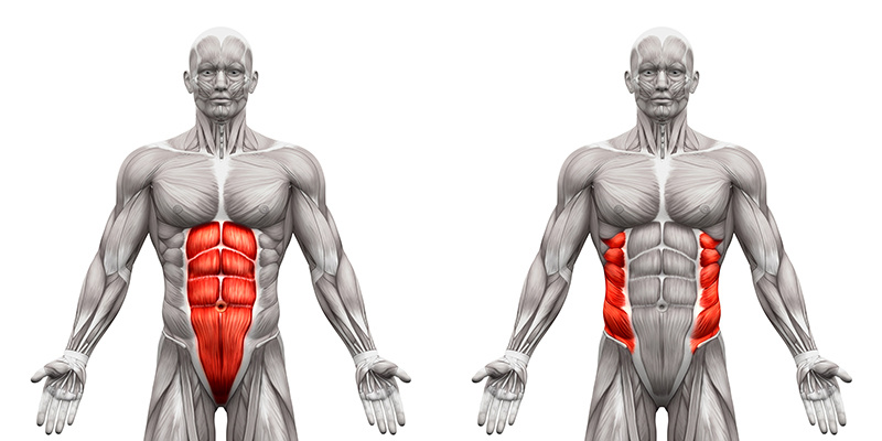
Abdominals refer to four muscles that are on the midsection, informally called abs by most bodybuilders. These include the rectus abdominis, the external obliques, the internal obliques, and the transverse abdominis. The best abdominal program uses exercises that target all four areas of the abdominal region—upper abs, lower abs, internal and external obliques, and the transverse abdominis.
The upper abs are best targeted with crunch exercises that involve flexing the upper spine forward by bringing the shoulders toward the hips, such as the standard crunch. The lower abdominals are best trained with exercises that involve flexing the lower spin forward by bringing the knees toward the chest, such as hanging knee raises.
Both the internal and external obliques are best targeted by exercises that flex the spine laterally to the left and right, such as oblique crunches. They also are targeted with exercises that involve flexing the spin forward and rotating it to the left or right, such as with crossover crunches.
The deep transverse abdominis is best trained with core exercises that fore the flexing of the transverse abdominis, pulling the navel in toward the spine to stabilize the spine and pelvis.
Abdominal Muscle Structure
Abdominal Muscle Function
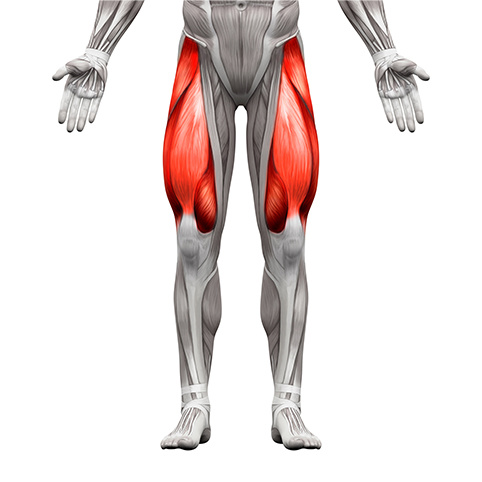
The quadriceps are the four muscles that make up the front of the thigh. The vastus lateralis, vastus medialis, vastus intermedius, and rectus femoris all originate from different attachment points on the thigh and hip bone, but they all converge on one common tendon to perform knee extension, such as when you kick a ball.
Because the rectus femoris originates on the hip bone, not the femur (thigh bone) as with the other three quadriceps muscles, it also is involved in hip flexion, such as when you lift your knee up.
Although all four muscles work together to straighten the knee, certain exercises are better for targeting specific parts of the quad. For instance, the leg extension best targets the rectus femoris muscle. However, doing leg extensions with the toes turned in places more stress on the outer quad (vastus lateralis), and doing leg extensions with the toes pointed out better targets the inner quads (vastus medialis).
The leg press hits all four quad muscles, but research shows that the emphasis is on the medialis muscle. Conversely, the hack squat tends to place more emphasis on the outer quads. Squats and lunges, however, hit the four quadriceps muscles fairly evenly, along with the leg adductors, hamstrings, gluteus maximus, and other muscles.
Quadriceps Muscle Structure
Quadriceps Muscle Function
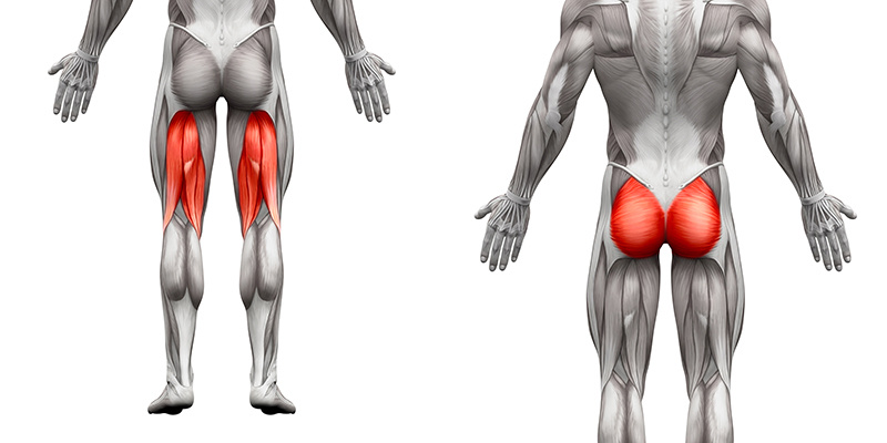
The hamstrings are the muscles on the back of the thigh. The gluteus maximus, also referred to as the glutes, are the large buttock muscles. The glutes are involved in extending the legs back, as when standing up from a seated position, and kicking the legs back behind the body. The hamstrings are composed of the biceps femoris, the semitendinosus, and the semimembranosus. Collectively the hamstring muscles not only flex the knee, as when you bend your knee, but they also work in conjunction with the glutes to extend the legs at the hips.
Although compound quadriceps exercises like the squat, hack squat, lunge, and step-up are traditionally considered quadriceps exercises, they also largely involve the glutes and the hamstring muscles. For this reason, most bodybuilders perform fewer hamstring exercises than quadriceps exercises.
Even though the hamstrings involve different muscles that work together to perform leg flexion and hip extension, specific exercises target each muscle. The Romanian Deadlift hits the entire hamstring fairly evenly along with the glutes because of the hip extension involved in the exercise. The biceps femoris is better targeted with lying and standing leg curls. The semitendinosus and semimembranosus, on the other hand, are better targeted with seated leg curls. Therefore, a thorough hamstring workout should include one exercise that involves hip extension and knee flexion.
Hamstring and Glute Muscle Structure
Hamstring and Glute Muscle Function
Common Hamstring and Glute Exercises
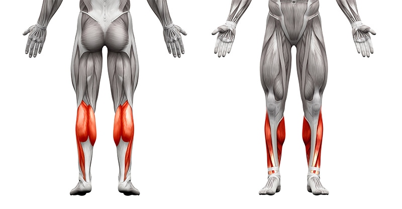
Calves refer to two separate muscles on the lower leg. These muscles are the gastrocnemius, a muscle shaped like an upside-down heart, and the soleus, a muscle that lies underneath the gastrocnemius. Both muscles perform extension at the ankle, such as when you stand up on your toes.
Certain exercises are better than others at targeting the two calf muscles. Standing calf raises, or any calf raise that involves a fairly straight knee, is better at focusing the stress to the gastrocnemius. The soleus, on the other hand, is better targeted with seated calf raises or any calf raise that is performed with the knee bent to about 90 degrees.
The best way to train calves is to include one or two exercises that target the gastrocnemius muscle and one exercise that targets the soleus muscle. Most bodybuilders train their calves after thighs. Some also include a second or third workout of the calves if they do not train legs twice a week. The reason for this is that the calves, particularly the soleus, are made up of a slightly higher percentage of slow-twitch muscle fibers. These muscle fibers have a high-endurance capacity and recover more quickly than fast-twitch muscle fibers. This Is also the reason that many bodybuilders train their calves with very high reps (20-30 per set). However, the best way to train calves is with the use of a periodized program that cycles the number of reps performed.
Calf Muscle Structure
Calf Muscle Function
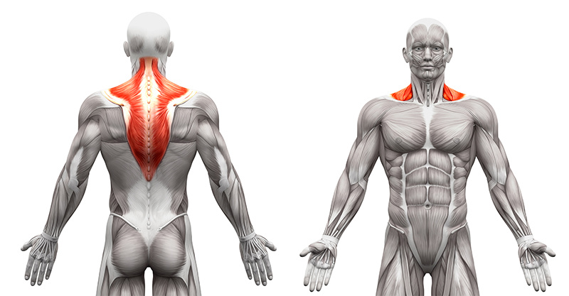
The trapezius is the large diamond-shaped muscle on the upper back, often referred to as traps. This muscle has upper, middle, and lower portions that all perform different movements. The upper trapezius primarily lifts and rotates the shoulder blades upward as when shrugging the shoulders, such as during dumbbell shrugs. The middle trapezius primarily pulls the shoulder blades together, such as during face pulls. The lower trapezius rotates the shoulder blades downward, such as when lifting a barbell overhead with straight arms like during the snatch.
Trapezius training can be paired with shoulders or back. Most bodybuilders train the traps after shoulders, because their primary interest is in developing the upper portion of the traps. The upper traps are involved in most deltoid exercises. Therefore, they are sufficiently warmed up after training shoulders.
However, because it is technically a back muscle and assists during many back exercises, upper traps are often trained with back. Most lifters typically pick one or two exercises for trap workouts and perform three to eight sets. If both a barbell and a dumbbell trap exercise are done in the same workout, the barbell exercise is typically done first.
Trapezius Muscle Structure
Trapezius Muscle Function
Common Trapezius Exercises
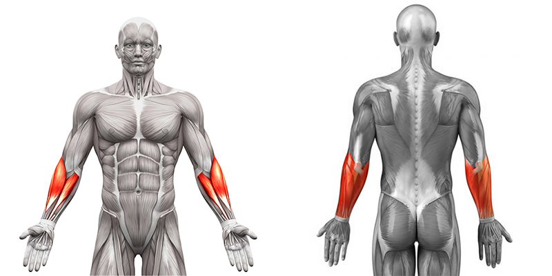
The forearms are the muscles that make up the entire lower arm. Though you do not need to familiarize yourself with all the different forearm muscles, you should recognize the difference in those referred to as the wrist flexor group and those referred to as the wrist extensor group.
The wrist flexor group is composed of forearm muscles that perform wrist flexion—the movement of the palms toward the inner forearm, such as during a wrist curl. The wrist extensors, on the other hand, are involved in performing wrist extension—moving the back of the hand toward the back of the forearm, such as when you twist the throttle on a motorcycle.
It is wise to train the forearms after the biceps because they are used so strongly during all biceps exercises. Typically, choosing one wrist curl or flexion exercise and one reverse wrist curl or extension exercise is sufficient for working the forearm muscles after biceps, especially if reverse-grip or hammer-grip curls were performed.
If grip strength is a limiting factor on back and biceps exercises, including a specific grip exercise may be warranted. The grip exercise should be done before the wrist curl and reverse wrist curl exercises. If training with a whole-body, upper- and lower-body, or two-day split, you may consider skipping specific forearm work and relying on the fact that the forearm muscles are used during back and biceps exercises.
Forearm Muscle Structure
Forearm Muscle Function
Common Forearm Exercises
Related Articles
Having well defined shoulders isnt easy...Having Boulder Sized Shoulders is difficult.
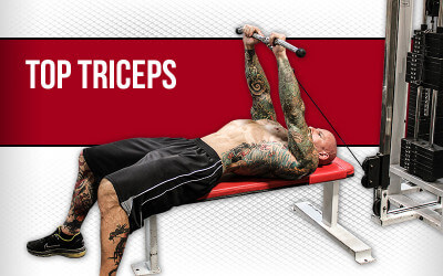
Keep your Triceps in TOP condition with this routine
More reps burns more calories? Maybe. The Doc Answers this and much More!!
Whats the Benefit of A-GPC? Find out exactly what it is.
Foundations
Featured Meal Plans
Muscle Hypertrophy: Build Muscle Fast
Rethink Your Rep Range
The Science of Strength Bands
Training
Nutrition
Supplementation
Health